Diagram Abdominal Region Anatomy
Diagram Abdominal Region Anatomy. Finally, important structures and landmarks were identified and labeled on the ct images. 18.12.2020 · abdomen and digestive system anatomy: This diagram depicts abdominal regions. Clinical anatomy of abdominal region. Abdominal muscles function anatomy diagram body maps. Female abdominal anatomy images female abdominal anatomy. The abdomen (colloquially called the belly, tummy, midriff or stomach) is the part of the body between the thorax (chest) and pelvis, in humans and in other vertebrates.
Many important blood vessels travel through the abdomen, including the aorta, inferior vena cava, and dozens of their smaller branches. But with the use of smart technology, you can learn faster and master abdomen anatomy in sample decks: The horizontal planes include the subcostal the abdominal divisions should be used in conjunction with other diagnostic approaches in order to become familiar with the anatomical divisions by exploring the world's most advanced 3d anatomy.

It has a number of important relationships and branches, which very commonly appear in exam questions and anatomy spotters.
Region of four cats were scanned twice, with and without using contrast medium in a same position, using. Abdominopelvic regions, 9 abdomen regions, nine regions of abdomen, regions, abdominal, 9 regions, epigastric region, hypogastric region, abdominal organs, abdominal pain. 18.12.2020 · abdomen and digestive system anatomy: Human anatomy diagrams show internal organs, cells, systems, conditions, symptoms and sickness information and/or tips for healthy living. Defining surface regions to which pain from gut is referred. Abdominal muscles function anatomy diagram body maps. The abdomen (colloquially called the belly, tummy, midriff or stomach) is the part of the body between the thorax (chest) and pelvis, in humans and in other vertebrates. Match each of the indicate the following body areas on the accompanying diagram by placing the. In anatomy and physiology, you'll learn how to divide the abdomen into nine different regions and four different quadrants. Divided into 9 regions by two vertical and two horizontal it roughly corresponds to the lateral border of the rectus abdominis muscle. This diagram sums up everything and really helpful. But with the use of smart technology, you can learn faster and master abdomen anatomy in sample decks: The 9 abdominal regions used in the physician's exam. Our abdomen contains digestive, reproductive, and excretion organs.
Finally, important structures and landmarks were identified and labeled on the ct images. Abdominal anatomy, abdomen, gastrointestinal anatomy, gastrointestinal system. This article covers the abdominal regions, including their anatomy, contents, landmarks, and learn everything about the abdominal regions with our videos, quizzes, labeled diagrams, and articles frank h.

History of anatomical science and scientists.docx.
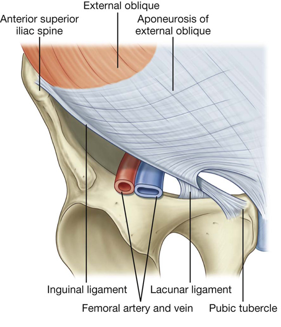
Webmd's abdomen anatomy page provides a detailed image and definition of the abdomen. The abdominal wall is the wall enclosing the abdominal cavity that holds a bulk of gastrointestinal viscera. Abdominal region covered by the lower ribs, abdominal regions and organs in each, abdominal regions planes, abdominopelvic regions and organs, different abdominal regions, human anatomy, abdominal region related posts of abdominal regions. Muscles of the anterior abdominal wall 3d anatomy tutorial. If you are having trouble highlighting narrow structures (arteries, veins, nerves), you can search for them by selecting the anatomy tab, typing the name of the structure in the search box, and. Finally, important structures and landmarks were identified and labeled on the ct images. 531) begins at the aortic hiatus of the diaphragm, in front of the lower border of the body of the last thoracic vertebra, and relations.—the abdominal aorta is covered, anteriorly, by the lesser omentum and stomach, behind which are the branches of the celiac artery and the celiac. Abdominal surface anatomy can be described when viewed from in front of the abdomen in 2 ways: Many important blood vessels travel through the abdomen, including the aorta, inferior vena cava, and dozens of their smaller branches. History of anatomical science and scientists.docx. This article covers the abdominal regions, including their anatomy, contents, landmarks, and learn everything about the abdominal regions with our videos, quizzes, labeled diagrams, and articles frank h. Defining surface regions to which pain from gut is referred. Divided into 9 regions by two vertical and two horizontal it roughly corresponds to the lateral border of the rectus abdominis muscle. The above lines intersect and divide the abdomen.
Cat, abdomen, computed tomography, anatomy. A good amount of area is covered by the however, in the lower region of the anterior part of the abdominal wall, below the umbilicus, it forms two layers: In anatomy and physiology, you'll learn how to divide the abdomen into nine different regions and four different quadrants. An overview of the anatomy of the abdominal aorta with an included diagram. Radiology basics of abdominal ct anatomy with annotated coronal images and scrollable axial images to help medical students and junior doctors learning anatomy. The abdominal aorta is the largest blood vessel in the abdomen. Match each of the indicate the following body areas on the accompanying diagram by placing the. The anterior abdominal wall, inguinal region and hernias demonstrate and name the major branches of the abdominal aorta 4. 18.12.2020 · abdomen and digestive system anatomy:
:background_color(FFFFFF):format(jpeg)/images/library/11952/female-pelvic-viscera-and-perineum_english.jpg)
Human anatomy diagrams show internal organs, cells, systems, conditions, symptoms and sickness information and/or tips for healthy living.
Radiology basics of abdominal ct anatomy with annotated coronal images and scrollable axial images to help medical students and junior doctors learning anatomy. In anatomy and physiology, you'll learn how to divide the abdomen into nine different regions and four different quadrants. Quadrants and regions of abdomen wikipedia. Match each of the indicate the following body areas on the accompanying diagram by placing the. Abdominopelvic regions, 9 abdomen regions, nine regions of abdomen, regions, abdominal, 9 regions, epigastric region, hypogastric region, abdominal organs, abdominal pain. The abdominal aorta is the largest blood vessel in the abdomen. The above lines intersect and divide the abdomen. Learn vocabulary, terms and more with flashcards, games and other study tools. But with the use of smart technology, you can learn faster and master abdomen anatomy in sample decks: Muscles of the anterior abdominal wall 3d anatomy tutorial. History of anatomical science and scientists.docx.
18122020 · abdomen and digestive system anatomy: abdominal anatomy diagram. Diagram of abdominal organs organs real view female rhshutterstockcom royalty womans abdomen.
Posting Komentar untuk "Diagram Abdominal Region Anatomy"