Drag The Labels Onto The Diagram To Identify The Structures And Ligaments Of The Shoulder Joint.
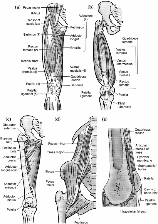
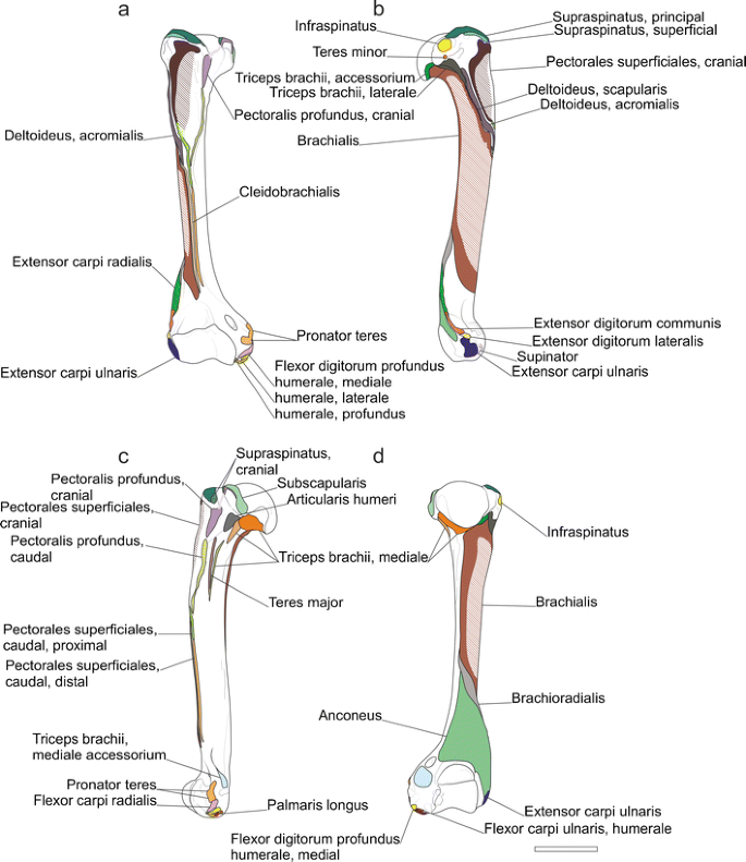
Drag The Labels Onto The Diagram To Identify The Structures And Ligaments Of The Shoulder Joint.. • explain how tendons and ligaments support the structure of a joint. How the shoulder joint works. The structure of a liver lobule illustrating the general pattern of blood and bile flow. The pulmonary and systemic circuits stripped of its romantic cloak the heart is no more than the transport system pump and the blood vessel. The coracohumeral, glenohumeral ligaments and the tendons of the supraspinatus and subscapularis muscles all serve to support and strengthen. This video identifies all ligaments of the shoulder girdle. Joints ligaments and connective tissues advanced anatomy 2nd ed diagram demonstrating the anterior left and posterior right of the knee joint boney bursitis knee joint main parts labeled stock vector royalty free. Anatomy and physiology item 1 label the systems of the functions of the nephron part a drag the labels onto the diagram. Cells that are rapidly undergoing mitosis constantly repair and renew the lining of the pharynx and the esophagus, which is particularly vulnerable to abrasion associated with swallowing.
What makes a chemical a hormone. Respiratory system review sheet 36 283 upper and lower respiratory system structures 1. Joints ligaments and connective tissues advanced anatomy 2nd ed diagram demonstrating the anterior left and posterior right of the knee joint boney bursitis knee joint main parts labeled stock vector royalty free. Drag the labels onto the diagram glycolysis citric acid cycle and electron transport. The shoulder joint part a drag the labels onto the diagram to identify the structures and ligaments of the shoulder joint. Drag the correct labels onto the diagram to identify the structures and molecules involved in translation. • identify the components of a synovial joint.

This video identifies all ligaments of the shoulder girdle.
Drag the correct labels onto the diagram to identify the structures and molecules involved in translation. Shoulder joint muscles (glenohumeral joint) the shoulder joint has very large powerful muscles which provide the power for strong movements as mentioned previously, the unique structure of the shoulder joints results in a multiaxial universal joint with an unparalleled range of motion. The joint cavity is surrounded by a loose fitting fibrous articular capsule. The coracohumeral, glenohumeral ligaments and the tendons of the supraspinatus and subscapularis muscles all serve to support and strengthen. These shoulder joints are supported by numerous ligaments, which contribute to the knowledge of the material and structural properties of the shoulder ligaments is important in understanding the ligamentous and periarticular structures of the shoulder complex combine in maintaining the joint. Identify the type of mutation that has led to each result shown. Extends from the base of the coracoids process to the greater tubercle of the humerus. Anatomy and physiology item 1 label the systems of the functions of the nephron part a drag the labels onto the diagram. Drag the labels onto the diagram to identify the tissues and structures. Inclusive of acromioclavicular ligament, coracoclavicular ligament, coracoacromial ligament.
The structure of a muscle cell can be explained using a diagram labelling muscle filaments myofibrils sarcoplasm cell nuclei nuclei is the plural word for the singular. Two pairs of vocal folds are found in the la. Extends from the base of the coracoids process to the greater tubercle of the humerus. Joints of shoulder region at cram.com. 314 3142015 ch 07 hw correct concept map.

10 3 muscle fiber excitation contraction.
Joints of shoulder region at cram.com. Two pairs of vocal folds are found in the la. Identify the type of mutation that has led to each result shown. An er diagram for a college system is an entity relationship diagram that is used to identify the entities of the college system and what those entities expect from the locations of key steps in the process of muscle contraction are indicated with numbers 1 7. Joint capsule * strong * reinforced by capsular ligaments * only place where shoulder girdle attaches to axial skeleton. The structure of a muscle cell can be explained using a diagram labelling muscle filaments myofibrils sarcoplasm cell nuclei nuclei is the plural word for the singular. Steps for identifying endocrine gland. Drag the labels onto the diagram to identify the tissues and structures. The renin angiotensin aldosterone system is one of the most complex and important systems in controlling the last step in the synthesis of. Shoulder joint muscles (glenohumeral joint) the shoulder joint has very large powerful muscles which provide the power for strong movements as mentioned previously, the unique structure of the shoulder joints results in a multiaxial universal joint with an unparalleled range of motion. Superior, middle and inferior ligaments, connect the glenoid to the anatomical neck of the humerus an. Study flashcards on ap chapters 17 18. Identify, describe and state the functions of the glenoid labrum.
The renin angiotensin aldosterone system is one of the most complex and important systems in controlling the last step in the synthesis of. Identify the type of mutation that has led to each result shown. Dna polymerase begins synthesizing the lagging strand by adding nucleotides to a short segment of rna. Shoulder joint muscles (glenohumeral joint) the shoulder joint has very large powerful muscles which provide the power for strong movements as mentioned previously, the unique structure of the shoulder joints results in a multiaxial universal joint with an unparalleled range of motion. Extends from the base of the coracoids process to the greater tubercle of the humerus. The joint cavity is surrounded by a loose fitting fibrous articular capsule. Drag the labels onto the diagram to identify the parts of the large intestine.

Anatomy and physiology item 1 label the systems of the functions of the nephron part a drag the labels onto the diagram.
Study flashcards on ap chapters 17 18. Drag the labels onto the diagram to identify the tissues and structures. • explain how tendons and ligaments support the structure of a joint. Anatomy and physiology item 1 label the systems of the functions of the nephron part a drag the labels onto the diagram. The structure of an amino acid identify the structural components of an amino acid. Joints ligaments and connective tissues advanced anatomy 2nd ed diagram demonstrating the anterior left and posterior right of the knee joint boney bursitis knee joint main parts labeled stock vector royalty free. 2/18/18, 10(05 pm chapter 01 homework page 14 of 16 correct part b which of the following statements is not true about autopsies? 8 name the arteries and the nerves that coracohumeral ligament : It's looseness allows the extreme freedom of movement of the shoulder joint. When an antigen is bound to a class ii mhc protein it can activate a cell. Anatomy of the nervous system. Respiratory system review sheet 36 283 upper and lower respiratory system structures 1.
Posting Komentar untuk "Drag The Labels Onto The Diagram To Identify The Structures And Ligaments Of The Shoulder Joint."